Modern exudate management: a review of wound
treatments
Author(s)
Richard White
PhD
Senior Research Fellow, Tissue Viability
Grampian NHS Trust,
Aberdeen, Scotland, UK.
Email: Richard@medicalwriter.co.uk
Keith F. CuttingPhD
Senior Research Fellow, Tissue Viability
Grampian NHS Trust,
Aberdeen, Scotland, UK.
Email: Richard@medicalwriter.co.uk
MN, RN, Dip N, Cert Ed
Principal Lecturer
Buckinghamshire Chilterns University College,
Chalfont St Giles, Buckinghamshire, UK.
Introduction
The management of wound exudate
requires the clinician to have an understanding of what it is, why it is
present and how to monitor and assess it accurately.
The production of wound exudate
occurs as a result of vasodilation during the early inflammatory stage of
healing under the influence of inflammatory mediators such as histamine and
bradykinin. It presents as serous fluid in the wound bed and is part of normal
wound healing in acute wounds. However, when the wound becomes ‘chronic’ and
non-healing with persistent, abnormal inflammation or when infection becomes
established, exudate takes on a different guise and generates clinical
challenges. In the chronic wound, exudate contains proteolytic enzymes and
other components not seen in acute wounds .
This type of exudate has justifiably been termed ‘a wounding agent in its own
right’ because it has the capacity to degrade growth factors and peri-wound
skin and predispose to inflammation .
In order to develop an effective management approach, the clinician must be
able to accurately assess and understand the implications of the composition
and quantity of exudate present in the wound.
Exudate
composition
Wound exudate was described by the
Swiss physician Paracelsus (c1491-1541) as nature’s balsam .
It is derived from serum through the inflammatory/extravasation process. Acute
wound exudate contains molecules and cells that are vital to support the
healing process. It has a high protein content (although lower than that found
in serum), with a specific gravity greater than 1.020. Its composition includes
electrolytes, glucose, cytokines, leukocytes, metalloproteinases, macrophages
and micro-organisms .
In the first 48 to 72 hours after wounding, platelets and fibrin may be
present, but this reduces as bleeding diminishes. See Table 1.
Component
|
Function
|
Fibrin
|
Clotting.
|
Platelets
|
Clotting.
|
Polymorphonuclearcytes
(PMNs)
|
Immune defence, production of
growth factors.
|
Lymphocytes
|
Immune defence.
|
Macrophages
|
Immune defence, production of
growth factors.
|
Micro-organisms
|
Exogenous factor.
|
Plasma proteins, albumin,
globulin, fibrinogen
|
Maintain osmotic pressure,
immunity, transport of macromolecules.
|
Lactic acid
|
|
Glucose
|
Cellular energy source.
|
Inorganic salts
|
Buffering, pH hydrogen ion
concentration in a solution.
|
Growth factors
|
Proteins controlling factor-specific
healing activities.
|
Wound debris/dead cells
|
No function.
|
Proteolytic enzymes
|
Enzymes that degrade protein,
including serine, cysteine, aspartic proteases and matrix metalloproteinases
(MMPs)
|
Tissue inhibitors of
metalloproteinases (TIMPS)
|
Controlled inhibition of
metalloproteinases.
|
As fluid passes through the inflamed
vessel walls (extravasation) it may be seen that wound exudate is in essence
modified serum and will therefore contain similar solutes. As it arrives at the
wound surface, this fluid may be contaminated with tissue debris and
micro-organisms.
Healing acute wounds produce exudate containing active growth factors. These
are not present in chronic wounds .
Appearance
of exudate
Modest amounts of thin, pale yellow
or straw-coloured exudate in an acute healing wound is considered normal. In
chronic wounds, the colour, consistency and amount of exudate may change as a
result of various physiological processes
See Table 2.
Type
|
Colour
|
Consistency
|
Significance
|
Serous
|
Clear, straw-coloured
|
Thin, watery
|
Normal. Possibly a sign of
infection. Some bacteria produce fibrinolysins, which degrade fibrin clots or
coagulated plasma. Some strains of Staphylococcus aureus, β-haemolytic
group A streptococci and Bacteroides fragilis, produce fibrinolysins. Pseudomonas
aeruginosa produces a non-specific enzyme that degrades fibrin.
|
Fibrinous
|
Cloudy
|
Thin
|
Contains fibrin protein
strands.
|
Serosanguinous
|
Clear, pink
|
Thin, watery
|
Normal.
|
Sanguinous
|
Red
|
Thin, watery
|
Trauma to blood vessels.
|
Seropurulent
|
Murky, yellow, cream-coffee
|
Thicker, creamy
|
Infection
|
Purulent
|
Yellow, grey, green
|
Thick
|
Infection. Contains pyogenic
organisms and other inflammatory cells.
|
Haemopurulent
|
Dark, blood-stained
|
Viscous, sticky
|
Contains neutrophils, dead/dying
bacteria and inflammatory cells. This means an established infection is
present. Consequent damage to dermal capillaries leads to blood
leakage.
|
Haemorrhagic
|
Red
|
Thick
|
Infection. Trauma. Capillaries are
so friable they readily break down and spontaneous bleeding occurs. Not to be
confused with bloody exudate produced by over-enthusiastic debridement.
|
[Adapted with permission from
Cutting KF. Exudate: composition and functions. In: White RJ, editor. Trends
in Wound Care Volume III. London: Quay Books, 2004 .]
Exudate
volume
In chronic wounds the inflammatory
response is altered owing to an uncontrolled expression of inflammatory
mediators with a concurrent increase in vascular permeability and the amount of
extravascular fluid. If the wound becomes infected, an abrupt increase in
exudate volume may be seen initially, followed by further quantitative and
qualitative changes. This has been attributed in part to specific bacterial
virulence mechanisms that result in vasodilation and extravasation.
Gautam et al (2001)
have described a process whereby neutrophils attracted to the site of injury
trigger the release of heparin-binding protein (HBP). It has also been shown
that chronic leg ulcer exudate contains increased levels of HBP when compared
to acute wound fluid .
It is likely that HBPs are implicated in the production of increased exudate.
Certain bacteria such as Pseudomonas aeruginosa stimulate the release of
HBP from neutrophils, thus aggravating chronic inflammation by augmenting
endothelial hyper-permeability .
Recent research has indicated that
some bacteria actually express histamine and thus, if present, produce an
additional physiological source of histamine in the wound environment. Morganella
species, for example M. morganii Gram-negative rods have been found to
express histamine .
Bacteria isolated from chronic wounds have been found to produce
physiologically significant levels of histamine.
It has yet to be determined if the production of this pro-inflammatory agent
may be effectively managed through the application of antihistamines.
Exudate volume
|
Effect
|
None
|
Wound tissues dry.
|
Scant
|
Wound tissues moist.
|
Small
|
Wound tissues wet; moisture evenly
distributed in wound; drainage involves 25% of dressing.
|
Moderate
|
Wound tissues saturated; drainage
may or may not be evenly distributed in wound; drainage involves 25-75% of
dressing.
|
Copious
|
Wound tissues bathed in fluid;
drainage freely expressed.
|
[Adapted with permission from
Bates-Jensen BM. The Pressure Sore Status Tool a few thousand assessments
later. Adv Wound Care 1997; 10(5): 65-73 .]
Exudate
assessment
Accurate assessment of the volume
and viscosity of exudate will indicate whether or not healing is progressing
normally.
Inspection of a dressing on removal
may yield valuable information on the level of exudate produced during dressing
wear time. To assess the exudate volume the healthcare practitioner should
count the number of dressings used over a time period, note the wear time for
individual dressings, examine the dressing for the presence of strikethrough
(wet or dry), examine the peri-wound skin condition and note any leakage .
Some of the subjectivity attached to
exudate assessment can be reduced by using a tool such as the exudate continuum
(Figure 1)
.
This is integral to the applied wound management approach described by Gray et
al .
This tool has the potential to assist in the accurate assessment of exudate and
lend support in the decision-making process. It offers a method of generating a
score relevant to volume and viscosity.
For example, if a score of 4 is
obtained using the exudate continuum (medium volume 3, and low viscosity 1) and
this increases to 8 (high volume 5, and medium viscosity 3) over three days
then the wound is likely to be deteriorating and may be infected. If the
intervention chosen is appropriate, for example an absorbent antimicrobial
dressing, then it is likely this will be reflected in a lower score after a few
days.
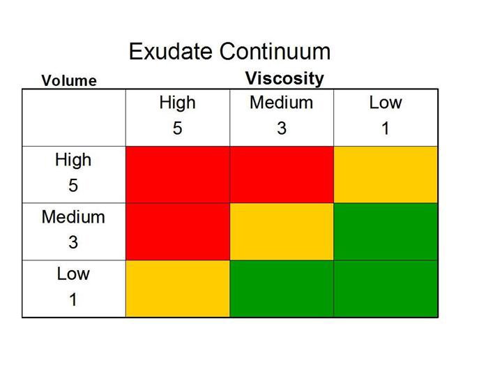
Figure 1 - The wound exudate continuum
[ Reproduced with permission from
Gray D, et al. Understanding applied wound management. Wounds UK
2005; 1(1): 62-8 .
]
Several wound management tools have
been developed that include a focus on exudate. One example is the wound bed
preparation model
with the development of the acronym TIME, in which ‘M’ represents the need to
maintain moisture balance .
An exudate management strategy has
been devised by Vowden and Vowden (2004).
This presents a strategy for assessing and evaluating the management of
exudate. See Table 4.
Cause
|
Control
|
Components
|
Containment
|
Correction
|
Complications
|
Systemic (local wound
related).
|
Whether effective systemic or
local control is possible
|
Bacterial load
Necrotic tissue
Chemical composition and pH
Viscosity and volume.
|
Dressing seal Where?
- at wound surface
- within dressing
- away from wound.
|
Modification of bacterial load
Debridement
Exudate modification.
|
Skin protection
Protein loss
Pain
Odour.
|
[Reproduced with permission from
Vowden K, Vowden P. The role of exudate in the healing process: understanding
exudate management. In: White RJ, editor. Trends in Wound Care Volume III.
London: Quay Books, 2004.
The selection of management options
should be based on the characteristics of the wound and the needs of the
patient. Successful management requires careful attention and continuous
evaluation throughout the lifetime of a wound.
For example, in the case of a leg ulcer, pressure ulcer or diabetic foot ulcer,
exudate levels in the non-infected wound are generally higher in the early
stages of healing and reduce as healing progresses. Dressing selection should
therefore be tailored to the condition of the wound. This might necessitate the
use of an absorbent moist dressing initially, changing to a moist dressing
suitable for low exudate levels at a later stage.
In wounds that become infected,
exudate levels often increase and exudate can become viscous. The focus here
should be on managing the underlying cause of infection.
Cavity wounds and other wounds left
to heal by secondary intention that are producing high levels of exudate may be
suitable for treatment with topical negative pressure. One method is vaccum
assisted closure™ (V.A.C® Therapy™), which can be effective in removing viscous
exudate.
Methods
used to manage exudate
If dressings are indicated, then
prudent selection and careful determination of wear time are imperative. This
will help ensure an optimal moist environment is maintained, while protecting
the surrounding skin from maceration.
Certain key performance
characteristics are required for any such dressing: they must absorb and retain
exudate, keep harmful chronic wound exudate away from the surrounding skin, perform
efficiently when used under compression, be easy to remove and be demonstrated
as cost-effective.
Wound dressings exhibit various
fluid-handling mechanisms: absorption, gelling, retention and moisture vapour
transmission. Information on a dressing’s fluid-handling mechanism is available
from the manufacturers. This information may not always be based on accepted,
independent test methodologies, but rather on in-house laboratory data, which
is invariably favourable to manufacturers’ own products. There are standard
test methods, published as monographs in various pharmacopoeias and in
peer-reviewed journals that provide independent, objective data on dressing
fluid handling .
The basic dressing mechanisms are as follows:
Absorption
Exudate is absorbed into the
dressing matrix. In the case of some foam dressings, this is a reversible
mechanism; the fluid can be expressed from the dressing under pressure. Not all
foams behave in this fashion.
Gelling
Following absorption, the exudate interacts
with the dressing material to form a gel .
This is a typical attribute of alginates: these carbohydrate polymers gel
according to the proportion of uronic acid units in their composition.
However, with alginate gels, fluid may come into contact with the peri-wound
skin .
This can also occur with hydrocolloid gel, the degree being dependent on
polymer composition.
Fluid
retention
In dressings with this mechanism,
fluid is absorbed by the dressing and is no longer available to wet the
surronding skin. Such materials retain the absorbed fluid directly above the
wound, without sideways spread or ‘lateral wicking’. An example of this is
Hydrofiber® technology. Such dressings have been demonstrated to be clinically
effective and cost-effective in exudate management, even when used under
compression.
Moisture
vapour transmission
In recent years dressings have been
designed to absorb fluid and, via an intermediate ‘wicking’ layer, move fluid
away from the wound/skin interface towards a permeable backing layer. Here,
some fluid is lost to the atmosphere by evaporation, a process known as
moisture vapour transmission. This mechanism is intended to increase the
fluid-handling capacity of the dressing .
The success of this process depends upon the proportion of absorbed fluid that
is lost. Evaporation will be compromised by the presence of occluding
materials, such as compression bandages, which may reduce evaporation rates.
There are no clinical data to suggest that this works in practice. Indeed, some
clinicians are sceptical that it has any performance-enhancing value.
Antimicrobial
properties
Dressings with an antimicrobial
component are intended for the control of the wound bioburden in critical
colonisation and local infection.
These dressings are useful, therefore, where raised exudate levels are
attributed to bacterial causes. There is also justification for their use in
cases of spreading infection where systemic antibiotics have been used and
impaired perfusion is suspected.
Typical antimicrobial dressings are those containing silver, iodine or honey.
Physical
therapies
Topical
negative pressure therapy
Suction drainage of wounds has been
used for many years,
and a variety of systems exist.
The integrated vaccum-assisted closure™ technique (V.A.C.® Therapy™) is claimed
to improve perfusion, reduce oedema and promote granulation tissue formation
and is supported by evidence from many wound types, including trauma wounds,
pressure ulcers, leg ulcers and surgical wounds.
The removal of exudate, particularly the more viscous forms, also removes
bacteria and protease enzymes – both barriers to healing. This technique
should, however, not be used on wounds containing eschar or necrotic tissue.
Compression
In the healthy limb, the return of
venous blood to the heart is achieved through the combined actions of the calf
muscle pump and the foot pump, both of which require reasonable mobility and
dorsiflexion of the ankle .
Where venous ulceration occurs, the patient needs assistance in achieving
venous blood return. Compression bandaging and intermittent pneumatic
compression therapy have both been found to be effective .
Whilst compression is recognised as the cornerstone of treatment for venous
disease, there are recognised limitations to current bandage systems, for
example the applied pressure is unknown, dependent upon method of application
and highly variable.
Compression therapy has two main
functions: to counteract venous hypertension and to control oedema.
In achieving these functions, exudate is reduced in the non-infected venous leg
ulcer.
In lymphoedema the application of appropriate compression bandages or garments
will result in a reduction both of the limb oedema and of any exudate leakage.
Intermittent pneumatic compression
(IPC) therapy is administered through a boot-shaped device which, by means of a
pump, is inflated and deflated to achieve alternating, dynamic compression of
the encased limb. IPC can be used as the main method of compression or as an
adjunct to orthodox compression bandaging.
Elevation/exercise
In venous leg ulceration, the
patient is advised to elevate the affected limb (with the ankle above the level
of the heart) to achieve venous blood return. While this may not always be
practical, some degree of elevation will aid venous return and, consequently,
reduce exudate. In lymphoedema, manual drainage
and exercise
are central to the control of oedema and leakage.
Conclusion
It is an unfortunate aspect of wound
management that exudate is often regarded as an issue only when it becomes a
clinical challenge - when leakage occurs or when peri-wound skin becomes
macerated.
However, such events are due to a combination of inaccurate assessment,
inappropriate dressing selection, over-optimistic wear time or poor patient
concordance. When dealing with purulent exudate, clinicians sometimes resort to
using absorbent pads, taped in position and changed frequently. While dressings
remain the mainstay of treatment, not all are suitable or effective for exudate
management.
Effective clinical management of
exuding wounds depends on accurate assessment of the volume and viscosity of
exudate, an understanding of relevant pathologies and the selection of an
appropriate exudate management mechanism. This is all too often left to a
dressing, without due consideration for other approaches.
A focus on this aspect of wound care
with the aim of developing clear recommendations for practice has the potential
to reduce morbidity and costs, and is therefore justified.
terimakasih ya informasinya...
ReplyDelete